Phytoplankton Removal Efficiency of an Experimental Sonication Technology
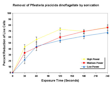
The goal of this project was to evaluate the efficiency of an experimental sonication technology in removing undesirable algal species from water samples, for potential use in treatment of water sources such as wastewater, drinking water, and ballast water. Three phytoplankton species encompassing a broad range in size (from ~4 mm to 100 mm along the major cell axis) were tested with this technology, including representative toxigenic, culturable cyanobacteria, diatoms, and dinoflagellates. Two of the taxa have resistant stages (cysts or akinetes) that were also tested with the sonication technology. The low-volume assay design included three levels of sonication intensity crossed with a minimum of four time intervals. Evaluation of these species enabled assessment of the efficacy of this sonication technology for removal of phytoplankton, including harmful species, as a general group.
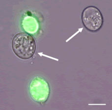
Fig 1. Light micrograph of sonicated Pfiesteria piscicida cysts treated with SYTOX Green, showing live cysts (arrows) and dead cysts (green fluorescence). Scale bar = 10 µm.
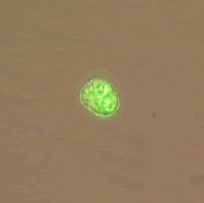
Fig 2. Light micrograph of Pfiesteria piscicida swimming cell after sonication at high power for one minute. Sonicated cells shed their flagella and formed immobile temporary cysts. Cells were stained with fluorescein diacetate (FDA). Live cells fluoresce green.
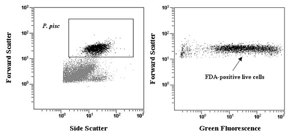
Fig 3. Histograms of flow cytometric viability assay of Pfiesteria piscicida after 30 seconds of high power sonication. Pfiesteria cells were detected by their light scatter signature, which was used to obtain a total cell count and to gate the histogram of green fluorescence. Cells in the region of positive FDA fluorescence were counted as live.
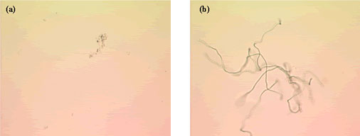
Fig 4. Light micrographs of (a) Anabaena flos-aquea cyanobacteria after sonication treatment at high power for four minutes, and (b) the same treatment following one week of re-culture under original culture conditions. All sonication power settings and exposure times up to four minutes showed vigorous re-growth, hence intact cells were counted as live.