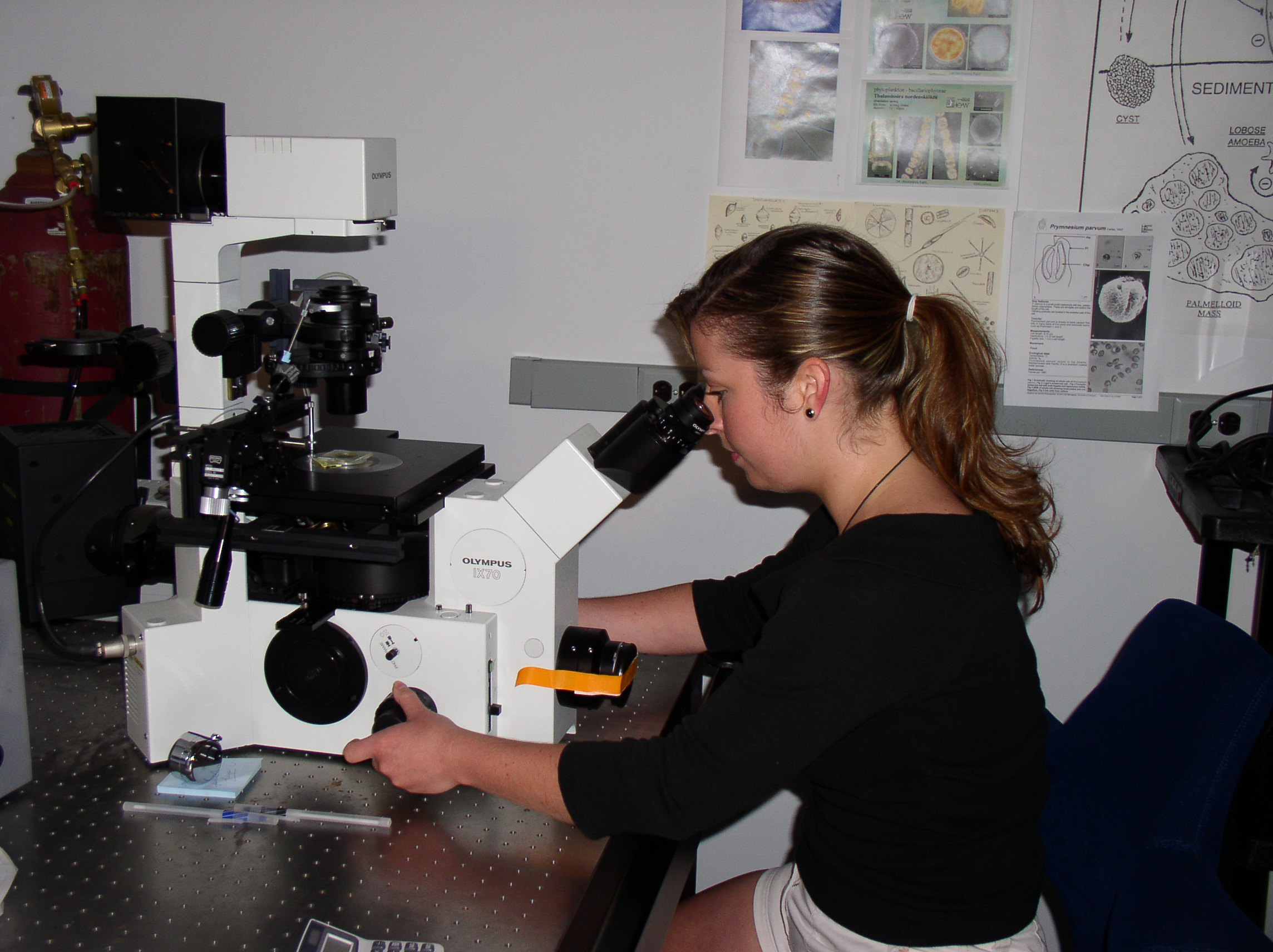
 Performing routine cell counts on various algal cultures under an Olympus IX70 inverted microscope.
Microscopy and Imaging Laboratory
The Microscopy and Imaging Laboratory is equipped with 5 research grade upright and inverted microscopes that use advanced fluorescence imaging technologies. These microscopes include an:
- Olympus Provis AX70 system equipped with a motorized x,y stage, water immersion objectives for live cell imaging, and capability for high-resolution fluorescence,Nomarski DIC, and phase contrast imaging;
- Olympus IX70 research inverted microscope with fluorescence and a micromanipulation package;
- Olympus CK40 inverted microscope for assessment of cell culture viability;
- Olympus SZX12 research stereomicroscope system with 0.3x and 1x objectives; and an
- Olympus BH-2 research microscope for “quick checks” and ease in quantifying cells.
Imaging systems capture both still and video images using an Olympus DP70 12.5 megapixel cooled digital color camera and an Optronics DEI-750T Peltier-cooled digital camera. These images are used in molecular, taxonomic and life cycle research of various microscopic organisms. The CAAE works in collaboration with the NCSU Center for Electron Microscopy and the Cellular and Molecular Imaging Facility for the most advanced scanning electron microscopy (SEM), transmission electron microscopy (TEM) and confocal microscopy available for biological research.
Example of still captures of Pfiesteria under a light microscope:
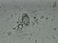
At the start of the clip, the zoospore begins to break through its protective shell.
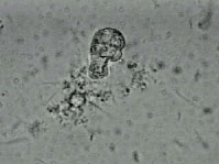
45 seconds later, the zoospore is mostly through the shell.
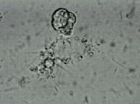
The zoospore emerges out of the shell.
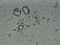
The zoospore has now completely reproduced.
